
Oxygen Nanobubble Delivery to Human Skin
Oxygenation, Barrier Integrity, and Adaptive Defense Response
Research led by Zvi Yaniv, PhD · Vivian Bucay, MD · Giuseppe Valacchi, PhD — NC State University
Watch Dr. Valacchi Present the Findings
In collaboration with North Carolina State University, AO2 SKIN tested whether topical oxygen nanobubbles could measurably improve oxygen availability in human skin. Using HIF-1α, the skin’s natural oxygen-response marker, researchers found a significant reduction after application, indicating improved oxygen delivery into the epidermis and dermis.
In this video, Dr. Giuseppe Valacchi of NC State University walks through the study design, methodology, data, and conclusions in detail.
Dr. Giuseppe Valacchi, PhD, NC State University · AO2 SKIN Clinical Science Presentation, 2026
Meet the Experts Behind the Study
This research was designed and conducted by three World-class doctors with deep expertise in dermatology, nanotechnology, and redox biology.

Dr. Zvi Yaniv, PhD

Vivian Bucay, MD, FAAD
Giuseppe Valacchi, PhD
Increase Skin Oxygenation, Proving it Biochemically
What we say it does. What the research shows.
Oxygen
A powerful antibacterial that is pure, natural, and the ideal opponent for anaerobic bacteria, the root cause of acne.Nanobubbles
Measured at one-billionth of a meter, they are 1,000× smaller than your skin pores, making them the ideal delivery vehicle.Concentration Gradient
High oxygen concentration in AO2 creates a dramatic gradient, allowing oxygenated nanobubbles to flood pores and clear anaerobic bacteria.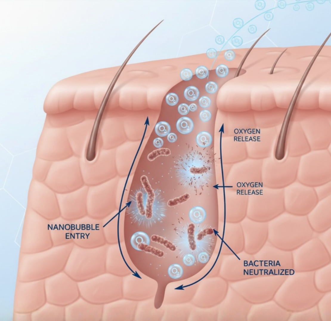
The Complex Structure of Skin and Its Oxygen Gradients
Human skin is not a uniform tissue, it maintains distinct oxygen tensions across the epidermis and dermis due to its layered anatomical structure. These gradients are measurable and, critically, measurably responsive to topical intervention. To validate that AO2 SKIN truly delivers oxygen into the tissue, not just onto the surface, the research team selected HIF-1α (Hypoxia-Inducible Factor 1-alpha) as the primary biomarker. This is the gold standard for measuring tissue oxygenation at the cellular level.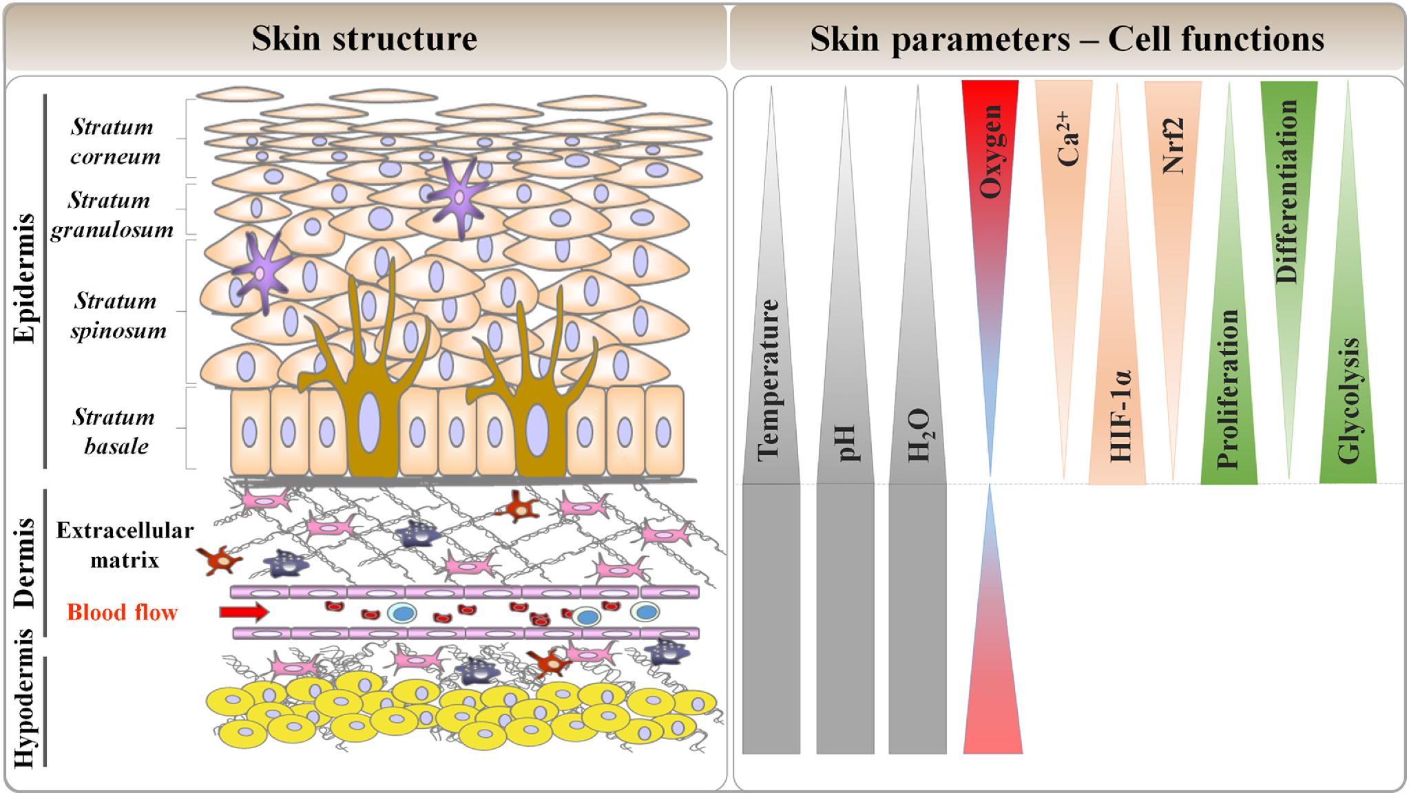
Oxygen tension gradients across skin layers (epidermis to dermis)

HIF-1α degradation pathway under normal vs. low oxygen conditions
HIF-1α: The Body’s Oxygen Alarm Signal
HIF-1α (Hypoxia-Inducible Factor 1-alpha) is the master transcription factor that regulates how cells respond to low oxygen levels. It is the most validated, widely-used indicator of tissue hypoxia in clinical research.
Under Normal Oxygen
HIF-1α is continuously degraded by the cell. No alarm signal is needed — the tissue is well-oxygenated.Under Low Oxygen (Hypoxia)
HIF-1α accumulates, translocates to the nucleus, binds to DNA, and activates stress-response genes.HIF-1α: The Body’s Oxygen Alarm Signal
HIF-1α (Hypoxia-Inducible Factor 1-alpha) is the master transcription factor that regulates how cells respond to low oxygen levels. It is the most validated, widely-used indicator of tissue hypoxia in clinical research.
Under Normal Oxygen
HIF-1α is continuously degraded by the cell. No alarm signal is needed, the tissue is well-oxygenated.Under Low Oxygen (Hypoxia)
HIF-1α accumulates, translocates to the nucleus, binds to DNA, and activates stress-response genes.
HIF-1α degradation pathway under normal vs. low oxygen conditions
Clinical Experimental Conditions
Biopsy collection protocol and anatomical site selection
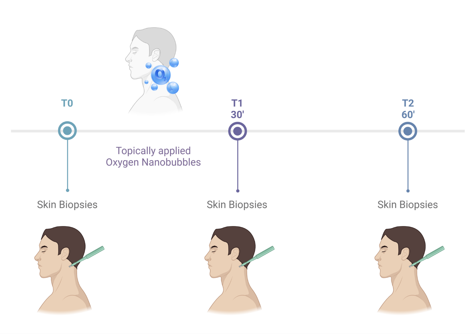
- Volunters: 5
- Time points: T0 (basal), T1 (30 min) and T2 (60 min)
- Treatment: Topically applied Oxygen Nanobubbles
- Skin samples: 3 biopsies (3mm) from the postauricular area
Study design:
Skin biopsies were collected from the postauricular area of five Volunteers, before and after treatment with topically applied Oxygen Nanobubbles for two points, i.e., 30 and 60 minutes after application. Skin biopsies will be processed for immunohistochemical analysis to evaluate the expression of HIF 1 alpha.
Study Results Overview
Confirmed
Confirmed
Confirmed
Confirmed
HIF-1α Decreases Significantly After Treatment
In untreated skin, HIF-1α (shown as red staining) was present at elevated levels, indicating the tissue was operating under relative hypoxia. After AO2 SKIN application, HIF-1α decreased significantly at both 30 and 60 minutes. The blue staining in the imagery serves only as a nuclear marker, confirming healthy, non-apoptotic cells are present throughout all time points.This is not a surface effect. The reduction in HIF-1α is a cellular response occurring inside the tissue, confirming that oxygen has been delivered across the skin barrier and into the dermal layers.
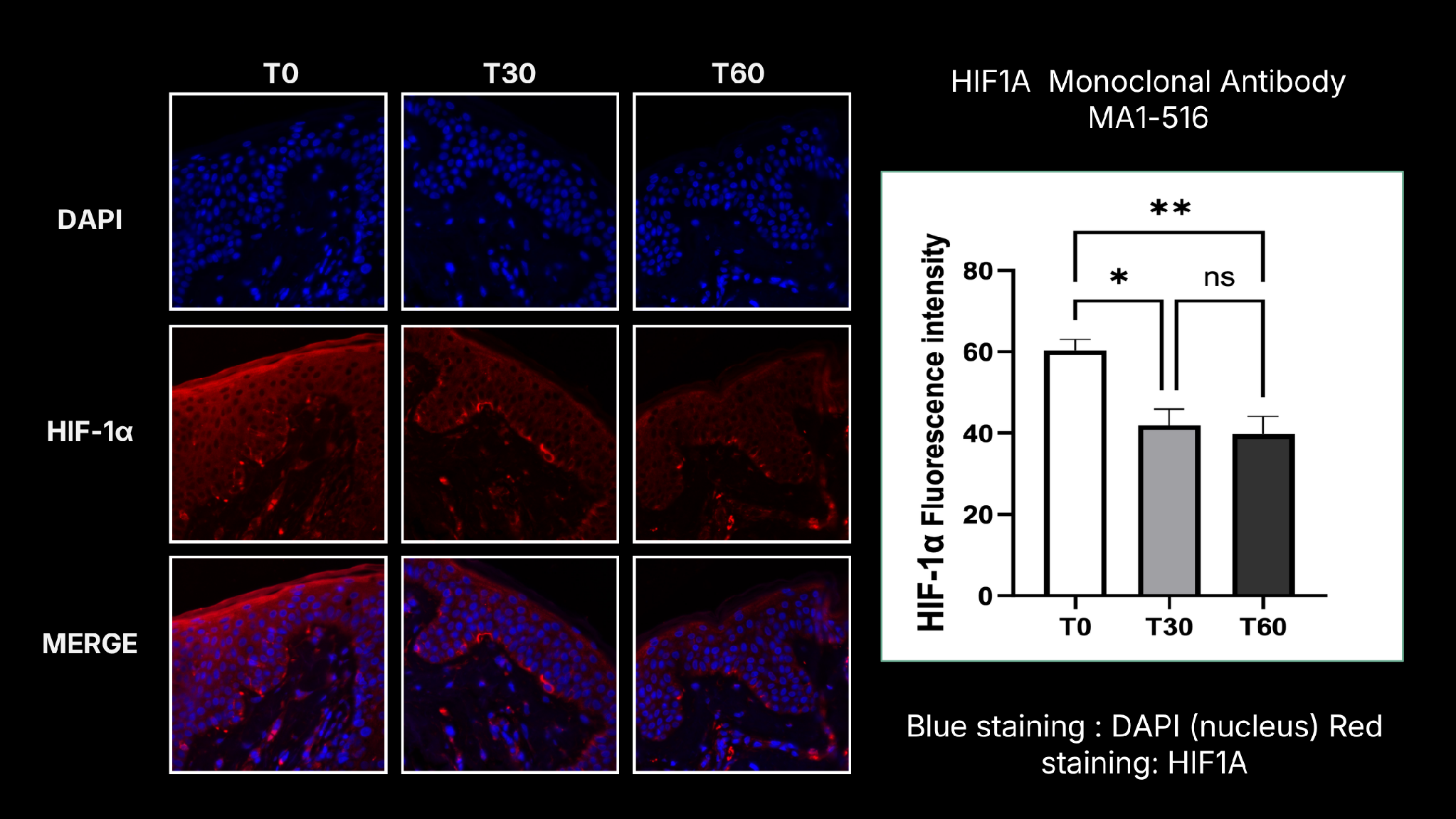
HIF-1α expression (red staining) at T0 (baseline), T30, and T60 minutes post-application. Significant reduction confirms oxygenation.
The Decrease in HIF-1α Confirms Effective Tissue Oxygenation
HIF-1α is a validated biomarker of tissue hypoxia. Its reduction after topical application is the gold standard confirmation that oxygen has been successfully delivered to the tissue using AO2 SKIN.
4-HNE: No Harmful Oxidative Stress Induced
Delivering additional oxygen into tissue raises an important clinical question: Does it create damaging oxidative stress? The data clearly answers no.
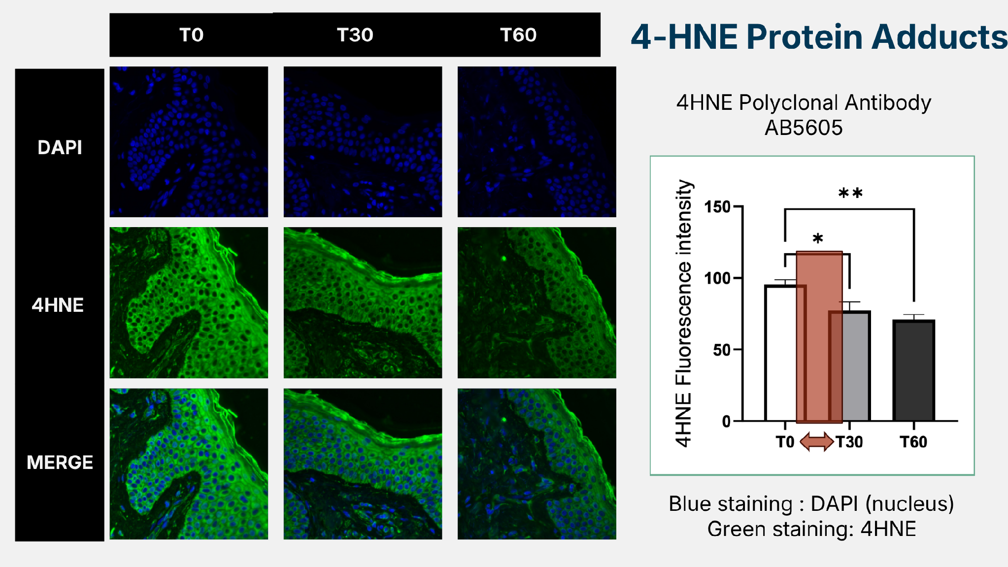
4-HNE (green staining) levels across treatment time points. No damaging increase detected.
About 4-HNE
4-HNE (4-Hydroxynonenal) is a stable marker of lipid peroxidation. At high concentrations, it forms protein adducts that cause cellular damage. At low levels, however, it functions as a beneficial signaling molecule that can activate the cell’s own defense pathways.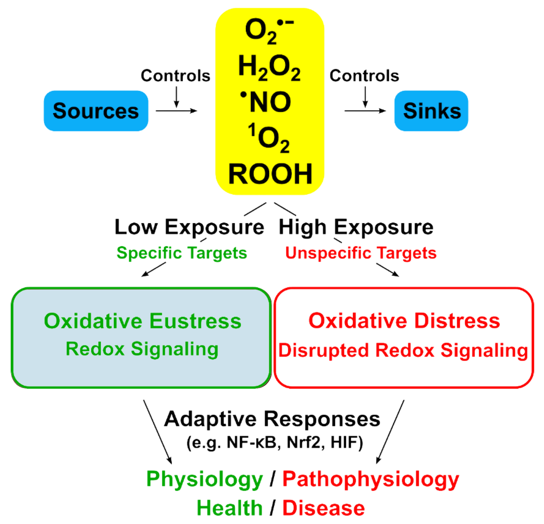
The hormetic dose-response: AO2 SKIN operates exclusively in the beneficial eustress range
Eustress vs. Distress: The Hormetic Principle
Not all oxidative signals are harmful. The dose makes the critical difference:
✓ Oxidative Eustress
Low-level, targeted signaling. Activates cellular defense pathways. Protective and physiological. This is where AO2 SKIN operates.✕ Oxidative Distress
High-level, untargeted damage. Disrupts redox signaling. Harmful to cells and tissue structure.AO2 SKIN Activates the Skin's Own Cellular Defense
The NRF2 pathway is the master switch for the skin's antioxidant defense system. When activated, it triggers downstream protective enzymes. The study measured all three components of this pathway.
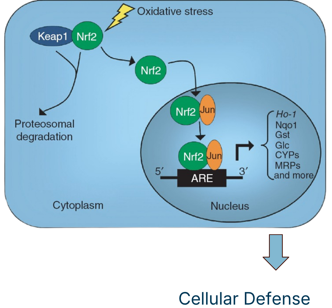
NRF2 → The Master Defense Transcription Factor
NRF2 is the sensor that detects oxidant signals and initiates the antioxidant cellular defense response. When activated, it migrates to the nucleus, binds to DNA, and transcribes key protective enzymes. An upward trend in NRF2 was observed, driving significant increases in its downstream targets.
Significantly Increased at T30
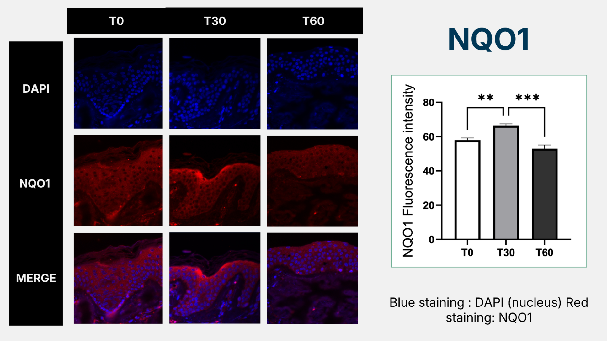 NQO1 is a protective enzyme regulated by NRF2. Its significant increase at 30 minutes confirms that AO2 SKIN activates the skin's own antioxidant defense system. Your skin becomes measurably stronger within 30 minutes of application.
Continues Rising Through T60, Sustained
 Unlike NQO1, HO-1 keeps increasing through the full 60-minute observation window. This suggests a sustained activation of cellular defense, protecting the skin from environmental stressors long after application.
Filaggrin Remains Completely Undisturbed
Filaggrin is the structural protein essential for skin barrier function and hydration regulation. It is incorporated into the lipid envelope of the stratum corneum and is a critical marker for epidermal homeostasis. To confirm the safety and hormetic (beneficial) nature of AO2 SKIN’s effects, the skin barrier structure must remain perfectly intact throughout treatment. Any disruption to filaggrin would indicate damage rather than benefit.This is clinically significant for skincare professionals: AO2 SKIN activates robust cellular defense responses while leaving the skin’s structural integrity completely undisturbed. The treatment is both effective and safe.
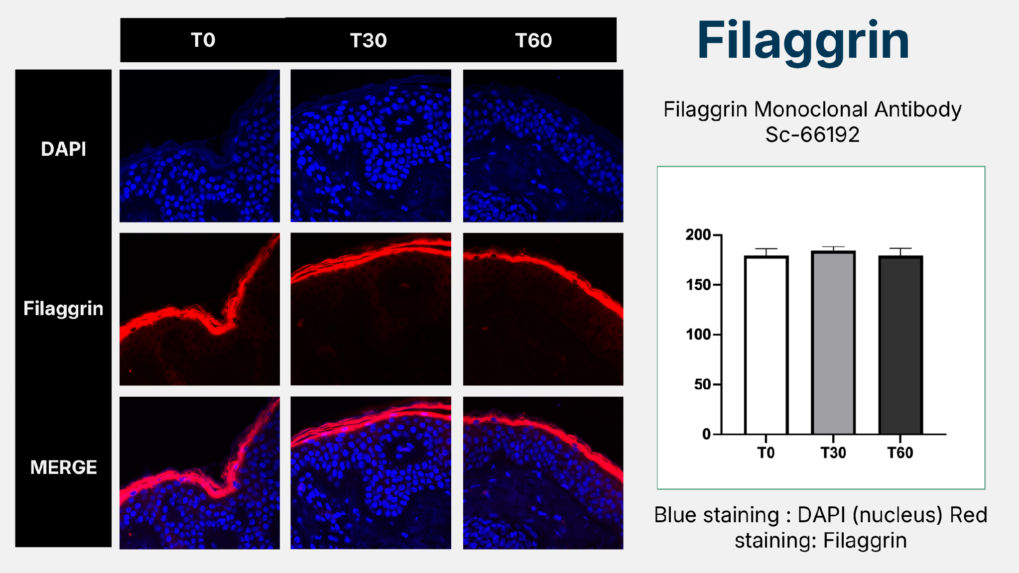
Filaggrin expression (red staining) remains stable across all time points. No disruption to skin barrier structure detected.
What the Science Confirms
Oxygen Delivery Confirmed
HIF-1α decreased significantly at both T30 and T60 providing direct biochemical evidence of improved tissue oxygenation. This is not anecdotal; it is measurable at the cellular level.
No Oxidative Distress, Only Eustress
4-HNE results confirm a transient, beneficial oxidative signal, not damaging stress. AO2 SKIN operates exclusively in the hormetic eustress range, where signals are protective, not harmful.
Cellular Defense System Activated
NQO1 was significantly elevated at T30. HO-1 continued rising through T60. The skin’s own antioxidant defense enzymes are measurably strengthened, and the effect is sustained.
Skin Barrier Fully Intact
Filaggrin levels remained completely unchanged across all time points. The treatment activates powerful cellular defense responses without any disruption to skin barrier structure or integrity.
“AO2 SKIN is able to deliver oxygen to the skin as measured by HIF-1α. The increase of oxygen does not induce oxidative distress, but most likely an oxidative eustress, the good one. The skin is much more able to defend itself from other stressors that could come after the application of this compound.”